
Urinalysis involves the physical, chemical, and microscopic examination of urine to detect various compounds and cellular components. It aids in diagnosing urinary tract infections, kidney diseases, and metabolic disorders.
Overview of Urinalysis
Urinalysis is a comprehensive diagnostic tool that involves the physical, chemical, and microscopic examination of urine to assess urinary tract health. It detects abnormal compounds, such as protein, glucose, and blood, and identifies cellular elements like red and white blood cells, epithelial cells, casts, and microorganisms. Microscopic examination plays a critical role in identifying structures like crystals, bacteria, and yeast, which are vital for diagnosing conditions such as urinary tract infections (UTIs), kidney diseases, and metabolic disorders. By analyzing these components, urinalysis provides valuable insights into the overall health of the urinary system and aids in early disease detection.
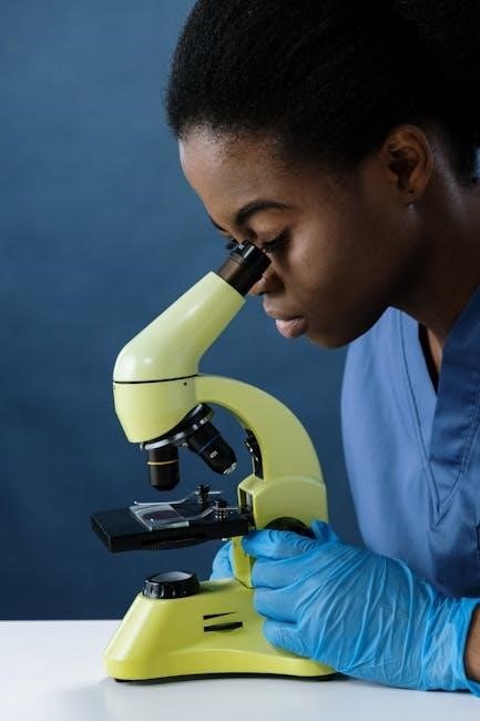
Importance of Microscopic Examination in Urinalysis
Microscopic examination is a cornerstone of urinalysis, providing critical insights into urinary tract health. It enables the detection of cellular elements, such as red and white blood cells, epithelial cells, casts, and microorganisms, which are essential for diagnosing conditions like urinary tract infections (UTIs), kidney diseases, and metabolic disorders. This method allows for the identification of structures that automated tests may miss, ensuring accurate and comprehensive diagnostic results. By revealing abnormalities in urine sediment, microscopic examination plays a vital role in early disease detection, guiding timely interventions and improving patient outcomes.

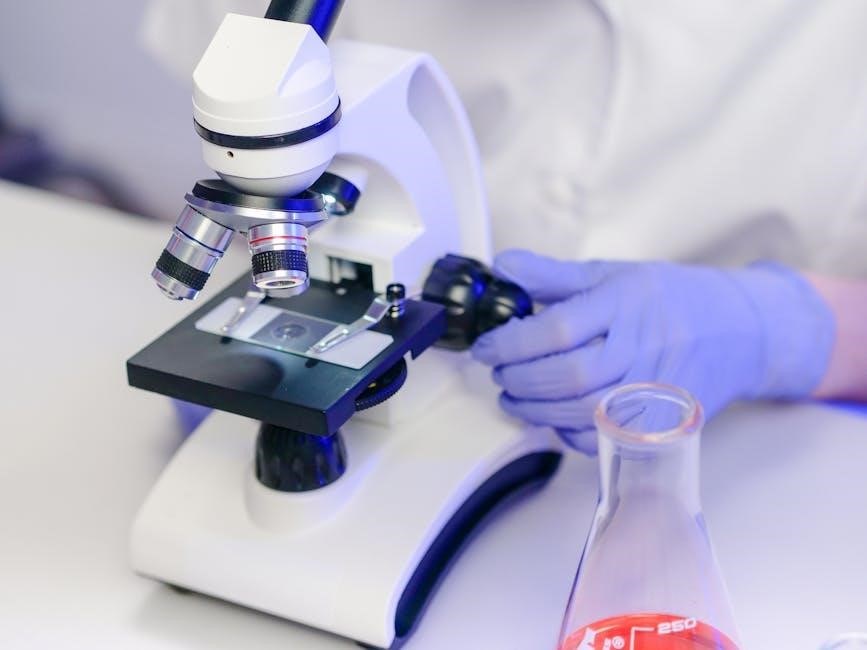
Methods of Urine Sample Preparation
Urine sample preparation involves collection, centrifugation, and slide preparation. Proper techniques ensure accurate microscopic analysis, preserving cellular components for reliable diagnostic results.
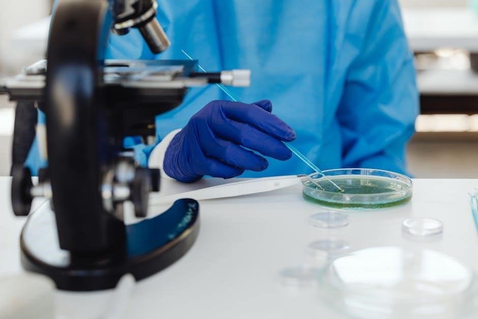
Collection and Centrifugation Techniques
Proper urine collection is crucial for accurate analysis. A midstream sample is preferred to minimize contamination. The sample is centrifuged at 2000 rpm for 5-10 minutes to separate the sediment, which contains cells, casts, and crystals. The supernatant is discarded, leaving a concentrated pellet for microscopic examination. This step ensures that cellular components are preserved for analysis. Proper centrifugation and handling techniques are essential to avoid sample degradation and ensure reliable diagnostic results. Standardized methods are followed to maintain consistency and accuracy in urine sample preparation.
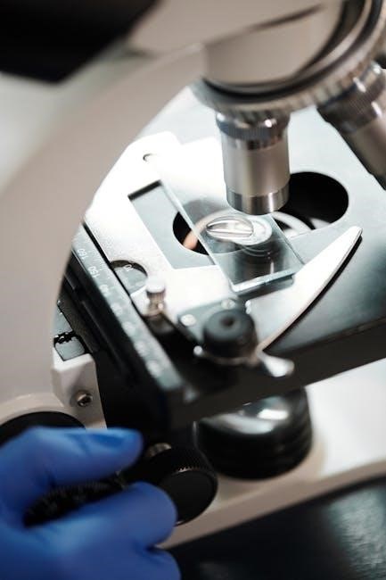
Slide Preparation for Microscopic Analysis
After centrifugation, a small amount of concentrated sediment is placed on a clean glass slide. A cover slip is carefully positioned over the sample to spread it evenly. This preparation allows for clear visualization under the microscope. Staining techniques, such as methylene blue or iodine, may be applied to enhance contrast and differentiate cellular components. Proper slide preparation ensures that all elements, including cells, casts, and crystals, are preserved for accurate microscopic examination. This step is critical for identifying abnormalities and diagnosing conditions effectively.

Components of Urine Sediment
Urine sediment contains various components, including red blood cells (RBCs), white blood cells (WBCs), epithelial cells, casts, crystals, and microorganisms, each providing diagnostic insights.
Red Blood Cells (RBCs) and Hematuria
Red blood cells (RBCs) in urine, known as hematuria, are a key finding in microscopic examination. Hematuria can be microscopic (visible only under a microscope) or gross (visible to the naked eye). The presence of RBCs may indicate urinary tract infections, kidney stones, glomerulonephritis, or other renal pathologies. RBCs are identified by their round, pale centers and pinkish-red color. The detection of RBCs, along with other sediment components, aids in diagnosing conditions such as inflammation, injury, or bleeding within the urinary system. Accurate identification and quantification of RBCs are crucial for clinical correlation and further diagnostic workup.
White Blood Cells (WBCs) and Leukocyturia
White blood cells (WBCs) in urine, referred to as leukocyturia, indicate the presence of inflammation or infection in the urinary tract. WBCs are large, granular cells with abundant cytoplasm and are often seen in conditions such as urinary tract infections (UTIs), pyelonephritis, or interstitial nephritis. Their presence, especially when accompanied by bacteria or other inflammatory markers, suggests an active immune response. Microscopic identification of WBCs is crucial for diagnosing infections and distinguishing them from other urinary abnormalities. The quantification of WBCs helps correlate findings with clinical conditions, guiding further diagnostic and therapeutic interventions. Leukocyturia is a key indicator of underlying urinary tract pathology.
Epithelial Cells and Their Significance
Epithelial cells in urine are derived from the lining of the urinary tract, including the kidneys, bladder, and urethra. Their presence can indicate various conditions, ranging from benign contamination to pathological states. Renal epithelial cells may suggest kidney damage, while transitional epithelial cells are common in normal urine. Squamous epithelial cells, often from the urethra or external genitalia, are frequently seen in contaminated samples. The type and quantity of epithelial cells help differentiate between physiological and pathological states, making their identification crucial for accurate diagnosis. Their significance lies in their role as markers of urinary tract health and potential disease.
Casts and Their Clinical Implications
Casts are cylindrical structures formed in renal tubules, composed of proteins and cellular elements. Their presence in urine sediment is a critical diagnostic marker. Hyaline casts are normal, especially after physical exertion, while granular casts suggest kidney disease or dehydration. Red blood cell casts indicate glomerulonephritis or inflammation, and white blood cell casts are linked to pyelonephritis. casts form when proteins and cells coalesce in concentrated urine within the renal tubules. Their type and quantity provide insights into kidney function and pathology, aiding in the diagnosis of various renal and urinary tract disorders. Accurate identification of casts is essential for targeted clinical management.
Crystals and Their Role in Diagnosis
Crystals in urine are solid particles formed from metabolic byproducts. Common types include calcium oxalate, uric acid, and phosphates. Their presence aids in diagnosing kidney stones, metabolic disorders, or renal dysfunction. Some crystals, like cystine, are rare and linked to genetic conditions. Crystal formation can occur in normal urine but may also indicate underlying disease. Microscopic examination identifies crystal types, helping differentiate between benign and pathological conditions. Accurate detection and classification of crystals are crucial for guiding clinical management and confirming diagnoses related to kidney function and metabolic health. Their presence often prompts further investigation to determine the cause of crystalluria.
Bacteria, Yeast, and Other Microorganisms
The presence of bacteria, yeast, or other microorganisms in urine is often indicative of a urinary tract infection (UTI). These pathogens can be identified during microscopic examination, where they appear as small, variably shaped structures. Bacteria such as E. coli are commonly observed, while yeast like Candida may also be present, especially in immunocompromised individuals. The detection of these microorganisms aids in diagnosing infections and guiding antimicrobial therapy. Microscopic findings are often correlated with urine culture results to confirm the diagnosis. Identifying these pathogens is crucial for targeted treatment and preventing complications in urinary tract infections. Accurate visualization under the microscope ensures reliable diagnostic outcomes.

Microscopic Examination Techniques
Microscopic techniques involve staining methods and centrifugation to enhance visibility of urine components. These methods aid in detecting cells, casts, and microorganisms, crucial for UTI diagnosis and analysis.
Staining Methods for Enhanced Visualization
Staining methods are essential for enhancing the visualization of urine components under a microscope. Common techniques include the use of methylene blue and safranin stains, which highlight cellular elements like red and white blood cells. These stains improve the contrast of structures such as casts, crystals, and microorganisms, making them easier to identify. Specialized staining protocols, such as Gram staining for bacteria, are also employed to detect infections. Additionally, some methods involve dyeing techniques to differentiate between various cell types and foreign particles. Proper staining ensures accurate identification of abnormalities, aiding in precise diagnosis and treatment planning for urinary tract infections and other conditions. This step is critical for reliable microscopic examination results.
Use of Microscopy in Detecting Urinary Tract Infections (UTIs)
Microscopy plays a pivotal role in detecting UTIs by identifying pathogens in urine samples. Under the microscope, the presence of bacteria, yeast, and white blood cells indicates infection. Microscopic examination allows for the detection of leukocyte esterase, a marker of inflammation. Additionally, the visualization of nitrites, produced by certain bacteria, further supports UTI diagnosis. Microscopy also helps in identifying casts and cellular debris associated with infections. Early detection through microscopy enables prompt treatment, reducing the risk of complications. This method remains a cornerstone in urinalysis for diagnosing and managing UTIs effectively.
Normal vs. Abnormal Findings
Normal urine typically contains few red or white blood cells, with no bacteria or casts. Abnormal findings include elevated cell counts, crystals, or microorganisms, indicating potential pathology.
Interpretation of Microscopic Results
Microscopic urinalysis results are interpreted by identifying abnormal components like red or white blood cells, casts, crystals, or microorganisms. The presence of red blood cells (RBCs) may indicate hematuria, while white blood cells (WBCs) suggest infection or inflammation. Casts, such as hyaline or granular casts, can signal kidney dysfunction. Crystals may point to metabolic disorders or kidney stone risk. Bacteria or yeast indicate potential urinary tract infections. Accurate interpretation requires correlating findings with clinical symptoms and patient history to avoid false positives or negatives, ensuring reliable diagnostic conclusions. Proper documentation and standardized terminology are essential for clear communication of results.
Correlation of Sediment Findings with Clinical Conditions
Correlating microscopic urine sediment findings with clinical conditions is essential for accurate diagnosis. The presence of red blood cells (RBCs) may indicate urinary tract infections or kidney stones, while white blood cells (WBCs) often suggest infections or inflammation. Casts can signal kidney dysfunction, and crystals may point to metabolic disorders. Bacteria or yeast indicate potential infections. Proper interpretation requires considering patient history and symptoms to avoid misdiagnosis. Accurate correlation ensures timely and appropriate treatment, highlighting the importance of skilled analysis in clinical practice.

Reporting Standards
Standardized terminology and clear documentation are essential in urinalysis reports. Findings must include specific elements like RBCs, WBCs, casts, crystals, and microorganisms, correlating with clinical conditions accurately.
Documentation of Microscopic Observations
Accurate documentation of microscopic findings is critical for reliable urinalysis results. Observations must include descriptions of cells, casts, crystals, and microorganisms, noting their quantity and characteristics. Standardized terminology ensures consistency across reports. Each finding should be clearly recorded, avoiding ambiguity. Proper documentation aids in tracking changes over time and correlating results with clinical conditions. Use of checklists or structured forms can enhance precision. Additionally, the date, patient identification, and sample details should accompany all reports to ensure traceability and accountability. Adherence to these standards ensures that microscopic observations are reliable and actionable for healthcare providers.
Standardized Terminology in Urinalysis Reports
Standardized terminology in urinalysis reports ensures clarity and consistency, enabling precise communication among healthcare professionals. Terms like “hematuria,” “leukocyturia,” and “crystaluria” are universally accepted. Uniform definitions for elements such as red blood cells, white blood cells, and casts are essential. This uniformity aids in accurate interpretation and reduces diagnostic errors. Laboratories worldwide adhere to these standards, facilitating data comparison and ensuring reliable patient care. Consistent terminology also supports the integration of manual and automated analysis systems. By maintaining standardized language, urinalysis reports contribute effectively to clinical decision-making and patient management. This approach is vital for maintaining high diagnostic accuracy and patient safety.

Clinical Applications
Microscopic urinalysis aids in diagnosing urinary tract infections, kidney diseases, and metabolic disorders. It is crucial for monitoring chronic conditions and assessing renal function in patients with underlying health issues.
Diagnosis of Kidney and Urinary Tract Disorders
Microscopic examination of urine is a cornerstone in diagnosing kidney and urinary tract disorders. It identifies red blood cells (RBCs), indicating hematuria, which may suggest kidney stones, glomerulonephritis, or urinary tract infections (UTIs). White blood cells (WBCs) point to leukocyturia, often associated with infections or inflammation. The presence of casts, such as granular or hyaline casts, can signal renal dysfunction. Additionally, crystals in urine may indicate metabolic disorders or kidney stone formation. This method also detects bacteria, yeast, and parasites, aiding in the diagnosis of UTIs and other infections. Early detection of these elements enables timely intervention, improving patient outcomes and disease management.
Monitoring of Chronic Conditions
Microscopic examination of urine plays a vital role in monitoring chronic conditions like diabetes, hypertension, and kidney disease. For diabetic patients, it helps detect glucose and ketones, indicating glycemic control. In hypertension, proteinuria (excess protein) is a marker of renal involvement. Regular urinalysis can also track kidney function in chronic kidney disease (CKD), identifying casts and protein levels. This allows for early adjustments in treatment, preventing disease progression. By providing insights into urinary abnormalities, microscopic analysis supports long-term management of these conditions, ensuring timely interventions and improved patient outcomes.
Microscopic examination of urine is crucial for detecting urinary tract infections, identifying cells, casts, and crystals, and aiding in the diagnosis of various clinical conditions.
Microscopic examination of urine is a critical diagnostic tool for identifying cellular components, casts, crystals, and microorganisms. It aids in detecting urinary tract infections, kidney diseases, and metabolic disorders. Key elements analyzed include red and white blood cells, epithelial cells, and bacteria. Staining techniques enhance visualization, while centrifugation concentrates sediment for accurate results. Standardized terminology ensures consistent reporting. This method is essential for correlating sediment findings with clinical conditions, enabling timely and precise diagnoses. Regular microscopic analysis in laboratories remains vital for comprehensive patient care and monitoring chronic conditions effectively.
Future Perspectives in Urine Microscopy
Advancements in technology are expected to revolutionize urine microscopy, with automation and AI-driven systems improving accuracy and efficiency. Digital microscopy and deep learning algorithms may enhance image analysis, enabling faster and more precise diagnostics. Point-of-care testing devices could simplify urine analysis, making it more accessible. Integration of molecular techniques, such as DNA analysis, could expand the diagnostic capabilities of urine microscopy. Additionally, the development of nanotechnology-based tools may improve the detection of biomarkers for early disease diagnosis. These innovations promise to make urine microscopy a more powerful and indispensable tool in clinical practice, fostering better patient outcomes and streamlined workflows.